QBARS - v31n3 Reflectance Spectrophotometric Analysis of Flower Color
Reflectance Spectrophotometric Analysis Of Flower Color
Bruce E. Bradley and Andrew Koran, III
Ann Arbor, Michigan
The most convenient and widely used method of comparing and communicating flower color is with the use of standardized charts. Of several commercially available forms, the one published by the Royal Horticultural Society appears to be the most commonly used system with regard to measuring rhododendron color.
The use of standardized charts for color measurement of flowers has several limitations, including the limited number of samples available, the variable introduced by the type and quality of light in which the chart is used, and the fact that, in the final analysis, it relies upon the subjective evaluation of the person making the comparison. All of these limitations are obviated by the measurement of color using the method of reflectance spectrophotometry.
Although reflectance spectrophotometric analysis is used as a tool in both science and industry, its application to the analysis of flower color has apparently been rather limited, especially in regard to the study of rhododendron. The purpose of this paper is: (1) to briefly describe the principals involved in the use of the instrument, (2) present examples of reflectance curves and data obtained from several varieties of rhododendron, and (3) suggest some possible avenues of research using this methodology which might be productive in furthering knowledge of the genus Rhododendron .
When light from any source strikes an object it can be either transmitted, absorbed, or reflected (or any combination of the three). Only reflected light reaches the eye and is, therefore, responsible for the visual sensation of color. In a reflectance spectrophotometer, the sample is placed in a port on a reflectance sphere and is exposed to known wavelengths of light from 400-700 nm (the visible spectrum). The energy which is reflected at intervals from 400-700 nm is measured and graphically recorded, giving a precise profile of the reflectance characteristics of the sample. The data from the graph is then analyzed by the use of a computer program. The program is based upon the CIE Chromaticity Diagram (1931).(Munsell Color, Baltimore, Md. 21218.)
The tri-stimulus values of color are computed and are used to determine the three parameters of color which are: (1) luminous reflectance, (2) dominant wavelength, and (3) excitation purity. Luminous reflectance is a measure of whiteness or blackness, with a value of 100 being pure white and 0 being pure black. It is similar to "value" used in visual systems of analyzing color. Dominant wavelength is the actual color perceived by the eye and is synonymous with "hue". Excitation purity is the amount of color present, with 1.0 being complete saturation of color (of a specific dominant wavelength) and 0 being complete absence of color. Excitation purity is synonymous with "chroma." These three parameters of color are independent of each other. When a spectrophotometer is used in a reflectance mode the color of any object that can be placed in the reflectance port can be determined with extreme accuracy.
Methods
Each flower was split to conform to a flat surface, wiped clean with a cotton pledget soaked in deionized water, and placed in the sample port. Curves of per cent reflectance versus wavelength (λ) were obtained for each sample between 400 and 700 nm with a double-beam, ultraviolet-visible spectrophotometer* and integrating sphere.** Samples were backed by a white BaSO4 standard*** and evaluated in the diffuse reflectance mode. A second white standard was used in a reference port for calibration and to obtain data. Scanning speed was 1 nm/sec with a bandwidth of 2 nm. Tri-stimulus values (X, Y, Z) relative to the 1931 CIE color-matching functions for CIE standard illuminant C were determined by numerical integration (Δ λ = 5 nm). Values of CIE chromaticity coordinates (x, y) were calculated from the tri-stimulus values and were used to obtain dominant wavelength and excitation purity. Luminous reflectance was equal to tri-stimulus value Y.
* ACTA CIII UV-Visible Spectrophotometer, Beckman Instruments, Inc., Irvine, CA.
**ASPH-U Integrating Sphere, Beckman Instruments, Inc., Irvine, CA
***Part No. 104384, Beckman Instruments, Inc., Irvine, CA
Results
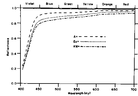
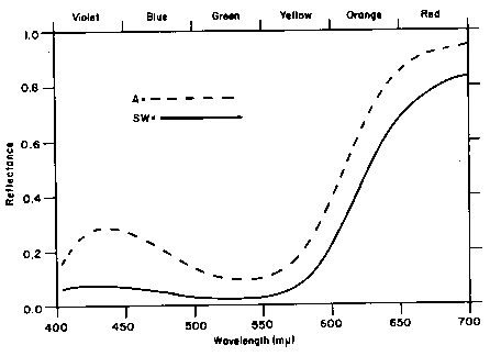
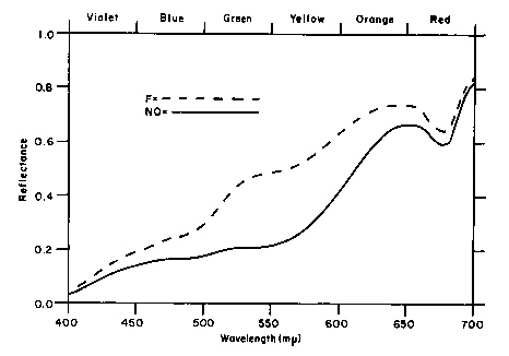
Reflectance curves are presented in Figures 1 through 3 to show, in a general way, comparisons of color between flowers and an example of what such data look like. More precise information can be derived from the table of numerical values of the tri-stimulus parameters. The designation of colors at the top of the reflectance curves is only a guide which correlates color to wavelength.
|
||
|
||
|
Figure 1 demonstrates the reflectance curves of three flowers from different clones of R. yakushimanum . (The straight line at the top of the figure represents the 100% calibration line and is the same for all figures.) A black standard is used for 0% calibration. All three flowers reflect nearly constant energy throughout the visible spectrum, thus appearing white to the eye. In this particular example, R . 'Angel' reflects the most energy. This is also seen in the table. 'Angel' has the highest luminous reflectance (95.36) of the three samples (100 = pure white, 0 = pure black). It also exhibits the lowest excitation purity (0.07), eg. compare to 'Scarlet Wonder' with an EP of 0.71, the highest EP of any sample tested. All three samples are very similar in dominant wavelength (583.42, 583.42, and 582.06) which is in the yellow portion of the visible spectrum.
Figure 2 compares two standard red rhododendron hybrids, 'America' and 'Scarlet Wonder'. Both flowers reflect strongly in the red portion of the spectrum. Note, however, the peak at about 440 nm in the curve from 'America'. The energy reflected in the violet portion of the visible spectrum accounts for the distinct magenta color of this variety.
The third figure compares two flowers from the hybrid 'Mary Belle'. The lower reflectance curve is of a newly opened flower which visually appears orange. The upper tracing is from an older, "faded" flower (which was structurally intact, i.e., showed no sign of wilting) which appeared to the eye as a pale yellow. Note, in the table, that the dominant wavelength has shifted from 600.19 to 588.64 nm. There is also a significant increase in luminous reflectance from 33.09 to 54.13. There is little change in excitation purity (0.57 to 0.59).
Discussion
The change in reflectance characteristics observed in 'Mary Belle' emphasizes a variable which might be difficult to standardize in analysis of rhododendron flower color.
It is apparent to most observers that the flowers on a truss of this variety change in color with time. This phenomenon is seen in many rhododendron flowers and it is likely that all flowers are slowly and continuously changing color from the time the bud begins to open until they wilt and drop from the plant. In the comparison of the three clones of R. yakushimanum , three flowers were selected which were subjectively judged to be "fully open" and showed no sign of pink coloration. The variation in reflectance curves among flowers from the same truss and of flowers from different trusses on the same plant, recorded at the same time interval after opening, should indicate this variability which must be recognized in any future comparative study.
| Table I | |||
|
Dominant
Wavelength |
Excitation
Purity |
Luminous
Reflectance |
|
| 1. R. yakushimanum 'Angel' | 582.06 | 0.07 | 95.36 |
| 2. R. yakushimanum Exbury | 583.02 | 0.11 | 91.96 |
| 3. R. yakushimanum F.C.C | 583.52 | 0.12 | 89.33 |
| 4. 'Scarlet Wonder' | 631.12 | 0.71 | 15.96 |
| 5. 'America' | -506.35* | 0.18 | 27.95 |
| 6. 'Mary Belle' (newly opened) | 600.19 | 0.57 | 33.09 |
| 7. 'Mary Belle' (faded) | 588.64 | 0.59 | 54.13 |
| * The negative sign indicates a complimentary wavelength and a dominant wavelength in the purple hue. | |||
Within the guidelines just mentioned, reflectance spectrophotometric analysis might be used to measure any variable which could potentially alter the color of rhododendron flowers. One of the more obvious variables might be the available concentrations of trace elements such as aluminum and iron, which are known to have a profound effect upon flower color.
Another possible application might be in the study of genetic transmittance of flower color. Such studies might be useful in suggesting crosses likely to achieve specific color in a hybridization program.
In conclusion, reflectance spectrophotometry adds an interesting and valuable dimension to research upon the genus rhododendron, but a note of caution can be given to those who would pursue it; this is, frustratingly, very seasonal research.
Reference
1. Kehr, August E., Research - What's New in '72, Qtr. Bull. A.R.S., Vol. 26, No. 4, 1972.


